Retained Root Removal for Tooth #36
Surgical Removal
Chief Complaint
Patient complains of pain in tooth #36.
Clinical Findings
Tooth #36 is very tender to percussion (TTP) and non-tender to palpation (NTTPp).
Mobility and periodontal depth are within normal limits (WNL).
Positive bite test.
Positive transillumination test.
Diagnosis of a cracked tooth with pulp involvement, resulting in pulpitis.
Treatment Plan
Extraction (XAP) with the possibility of oral surgery (OP) if complications arise.
Root canal treatment (RCT) followed by a post and crown (PC).
Pre-operative Assessment
Pre-operative CBCT scan taken to assess the condition and plan the procedure.
Anesthesia
Local anesthesia (LA) administered for the extraction (XAP) of tooth #36.
Surgical Procedure
Patient was cleaned and draped.
Incision made and buccal flap raised.
Bone guttering performed.
Attempted extraction revealed very firm roots deeply retained in the socket, with difficult access due to very dense bone.
Unable to successfully remove retained roots through initial extraction.
Proceed with oral surgery for the removal of retained mesial and distal roots of tooth #36.
Local anesthesia increased for the oral surgery.
Patient was cleaned and draped.
Incision made and buccal flap raised.
Bone guttering performed.
Mesial and distal roots of tooth #36 elevated out in two pieces.
Currettage performed and the site was irrigated with saline.
Four BSS (bone substitute materials) placed for primary closure.
Hemostasis achieved, and post-operative instructions (POI) were provided.


Post-operative Assessment
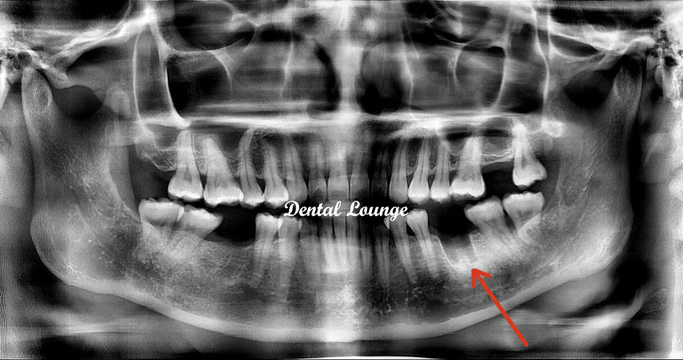
A post-operative CBCT scan confirmed that the inferior dental nerve (IDN) was spared and no remnants of the tooth were noted.
Patient informed and reassured.